1. Cell structure and Function
1.7. Cellular Functions
Cellular Functions
Dr V Malathi and Mrs Sushumna Rao
The basic functions of the cell include:
- Movement of substances across cell membrane,
- Cell division
- Protein Synthesis
I.Movement of substances across the cell membrane
The cell maintains a difference in the concentration of material between its exterior and interior . This is very important for the cell survival. The plasma membrane of the cell plays an active role in regulating the concentration of substances between the cell interior and exterior .
The plasma membranes are selectively permeable i.e., they allow some substances to pass through, but not others.
The main mechanisms of movement across cell membrane include:
Passive transport : is a naturally occurring phenomenon . The cell does not require energy to accomplish this movement. In this mode of transport substances move from an area of higher concentration to an area of lower concentration. A physical space in which there is a range of concentrations of a single substance is said to have a concentration gradient.
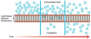
Diffusion : Nonpolar molecules, such as Oxygen, Carbon dioxide and Nitrogen
can move across the membrane from a higher concentration region to a lower concentration region through the process of diffusion. Diffusion does not require energy and hence this is called passive transport.

“Simple Diffusion across Plasma membrane” is licensed under CC BY 3.0
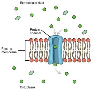
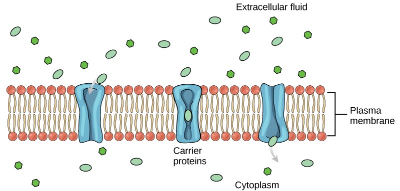
Facilitated diffusion : In this type of movement membrane proteins helps in the transport of materials across plasma membrane . The existing concentration gradient exists allow these materials to diffuse into the cell without expending cellular energy. However, these materials are ions or polar molecules and hence are repelled by the hydrophobic parts of the cell membrane. Facilitated diffusion proteins called transport proteins , shield these materials from the repulsive force of the membrane and helps them to diffuse into the cell. The transport proteins and can be channels or carrier proteins.
Channel proteins : These are transmembrane proteins. These proteins fold forming a channel or pore through the membrane specific for one particular substance. These proteins have hydrophilic domains exposed to the intracellular and extracellular fluids and also have a hydrophilic channel through their core that provides a hydrophilic opening through the membrane layers. Polar compounds can easily pass through the channel avoiding the nonpolar central layer of the plasma membrane that would otherwise slow or prevent their entry into the cell. Aquaporins are channel proteins that allow water to pass through the membrane at a very high rate.

“Facilitated transport” by Mariana Ruiz Villareal (modified ) is licensed under CC BY-NC 4.0
Carrier Proteins : Like channels, carrier proteins are trans membrane proteins, usually specific for particular molecules. These bind a substance and, in the process, trigger a change of its own shape, moving the bound molecule across the membrane. Large molecules which cannot pass through channels, such as amino acids and glucose are transported using Carrier proteins.
 “Facilitated transport” by Mariana Ruiz Villareal (modified ) is licensed under CC BY-NC 4.0
Osmosis : Osmosis is the diffusion of water across a semipermeable membrane. This diffusion depends on the concentration gradient, or the amount of water on each side of the membrane. The amount of water is inversely proportional to the concentration of solutes i.e., the higher the concentration of water, the lower the concentration of solutes, and vice versa. Due to the presence of aquaporins water can move readily across most membranes but the membrane limits the diffusion of solutes in the water.

“Osmosis” by OpenStax is licensed under CC BY 4.0
Tonicity describes how an extracellular solution can change the volume of a cell by affecting osmosis. To learn more about tonicity click on the link Tonicity
Active Transport: This mechanism of transport requires energy, usually in the form of adenosine triphosphate (ATP) and transports substances against concentration gradient that is, if the concentration of the substance inside the cell is greater than its concentration in the extracellular fluid (and vice versa). Active transport mechanisms move both small-molecular weight materials, such as ions, through the membrane as well as larger molecules.
Proteins for Active Transport : Specific proteins called transporters facilitate active transport. There are three types of transporters.
These are of 3 types namely
Uniporter : carries one specific ion or molecule.
Symporter : carries two different ions or molecules, both in the same direction.
Antiporter : carries two different ions or molecules in different directions.

“Active Transport Proteins” by Connectivid-D via Wikimedia Commons is licensed under CC BY-SA 4.0
Endocytosis : This is a type of active transport. Large molecules, whole cells and cellular parts are moved into a cell by this process. The steps in endocytosis are:
- The plasma membrane of the cell invaginates, forming a pocket around the target particle.
- The pocket pinches off, with the particle of transport being contained in a newly created intracellular vesicle formed from the plasma membrane.
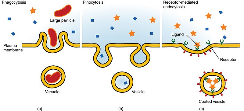
The three types of endocytosis are phagocytosis, pinocytosis, and receptor-mediated endocytosis.
Phagocytosis : This is a process of “cell eating” . For example , Large particles, such as other cells or microorganisms when invade the human body the neutrophil will “eat” the invaders through phagocytosis. The neutophils surround and engulf the microorganism, which is then destroyed by lysosomes inside the neutrophil .
The basic steps of phagocytosis include:
- The the inward-facing surface of the plasma membrane is coated with a protein called clathrin,
- The coated portion of the membrane then extends from the body of the cell and surrounds the particle, eventually enclosing it.
- The clathrin then disengages from the membrane
- The vesicle then merges with a lysosome and enclosed substance is broken down
- Nutrients from the degradation of the vesicular contents are extracted,
- The newly formed endosome merges with the plasma membrane and releases its contents into the extracellular fluid. The endosomal membrane again becomes part of the plasma membrane.
Pinocytosis : This is the process of “cell drinking”. Cells take in molecules, including water, which the cell needs from the extracellular fluid. This process results in a much smaller vesicle than phagocytosis, and they do not need to merge with a lysosome.
Receptor-mediated endocytosis : is a type of endocytosis that employs receptor proteins in the plasma membrane that have a specific binding affinity for certain substances. Clathrin protein attached to the cytoplasmic side of the plasma membrane are used for the process.
For example, the Low density lipoprotein also referred to as “bad” cholesterol is removed from the blood by receptor-mediated endocytosis.
In the human genetic disease familial hypercholesterolemia, the LDL receptors are defective or missing entirely. People with this condition have increased levels of cholesterol in their blood as their cells cannot clear LDL particles from their blood.
Although receptor-mediated endocytosis is designed to bring specific substances that are normally found in the extracellular fluid into the cell, other substances may gain entry into the cell at the same site.
Flu viruses, diphtheria, and cholera toxin all have sites that cross-react with normal receptor-binding sites and gain entry into cells.
 “Three Forms of Endocytosis” by OpenStax, is licensed under CC BY 4.0
Exocytosis : This process involves the moving of material out of the cell .The purpose of exocytosis is to expel material from the cell into the extracellular fluid.
Usually waste material is enveloped in vesicle and expelled into the extracellular space .
Proteins such as hormones, neurotransmitters, or parts of the extracellular matrix are also secreted by the cell through the process of exocytosis.

“Exocytosis” by OpenStax is licensed under CC BY 4.0
II. Cell division
The new cells required for the process of growth , repair and replacement are formed by the process of cell division.
The process of cell division includes two phases namely
(i) Cytokinesis : division of the cytoplasm and
(2) Karyokinesis : Division of the nucleus
There are two main types of cell division namely
Mitosis : This is the process by which the cells of the body , namely somatic cells divides. In this process two cells identical to the parent are produced

“Mitosis “ by Schoolbag.info is licensed under CC BY-SA 4.0
Meiosis : This is the process of cell division of the gametic cells ( Cells that give rise to sperm and ova). This process is also called reduction division as this division produces 4 cells with half the number of chromosomes than the parent cell.

“Mitosis Vs Meiosis” by Community College Consortium for Bioscience Credentials, is licensed under CC BY 3.0
III .DNA Replication and Protein synthesis
DNA Replication
Deoxyribonucleic acid (abbreviated DNA) is the molecule that carries genetic information for the development and functioning of an organism.
DNA has a double helical structure i.e., it is made of two linked strands that wind around each other to resemble a twisted ladder
Each strand of DNA has a backbone made of alternating sugar (deoxyribose) and phosphate groups.
Each sugar is attached to one of four bases: adenine (A), cytosine (C), guanine (G) or thymine (T).
The two strands of the DNA are connected by chemical bonds between the bases: adenine bonds with thymine, and cytosine bonds with guanine.
The sequence of the bases along DNA’s backbone encodes biological information for the synthesis of a protein or RNA molecule.
The DNA must be replicated once before every cell division.
Protein Synthesis
Yet another important function of the cell is protein synthesis.
Proteins serve as both structural and functional elements of the cell.
The synthesis of proteins consumes more of a cell’s energy than any other metabolic process.
Proteins account for more mass than any other macromolecule of living organisms.
The molecular process of protein synthesis, is called Translation .
it is the second part of gene expression and involves the decoding of the m RNA by a ribosome into a polypeptide product.
These are channel proteins that allow water to pass through the membrane at very high rate
The process of cell eating
Process of cell drinking
The process of moving material out of the cell
Division of cytoplasm
Division of the nucleus
Process by which the somatic cells divide
The process of cell division of the gametic cells
The molecular process of protein synthesis

