1. Cell structure and Function
1.4.Animal cell
Animal cell
Dr V Malathi and Mrs Sushumna Rao
Animal cells are the fundamental units of animal tissues and organs.
They are eukaryotic cells and therefore have membrane-bound organelles suspended in the cytoplasm enveloped by a plasma membrane.
Animal cells have centrosomes (or a pair of centrioles), and lysosomes which the plant cells do not possess.
A typical structure of an animal cell includes Organelles ( We will discuss about this in chapter 1.5) , cytoplasmic structures, Cytosol and Cell membrane .
The cell organelles like Centrosomes and lysosomes are found only in animal cells, but do not exist within plant cells
“Animal cell “ by OpenStax,via Wiki media commons is licensed under CC BY 4.0
Cell Membrane or the Plasma membrane
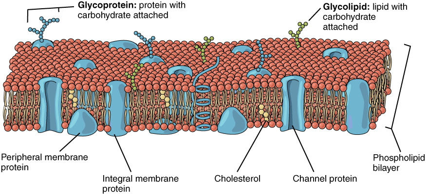
The cell membrane of an animal cell is a lipid bilayer which is embedded with other molecules like proteins, cholesterol and with some carbohydrates attached.
It separates the interior of the cell from the outside environment.
The cell membrane is semipermeable. The cell membrane regulates the transport of materials entering and exiting the cell.
The plasma membrane as it is other wise called permits the entry of small non polar molecules with ease, while large , polar molecules requires the help of transporters like membrane proteins .
The cholesterol embedded in the membrane provides structural integrity and support. Furthermore, the presence of cholesterol makes the membrane fluid rather than rigid, and renders them the capability of movement.

“Cell membrane “ by Openstax is licensed under CC BY 4.0
The membrane proteins are of two categories namely Integral membrane proteins and peripheral membrane proteins
Integral membrane proteins
These are otherwise called as Intrinsic proteins . These are proteins that are permanently attached to the cell membrane.
Their functions include : channeling or transport molecules across the membrane , cell receptors, cell adhesion
They are further classified as
- Transmembrane proteins : these span the entire plasma membrane and are found in all types of biological membranes
- Integral monotypic proteins : these are permanently attached to the membrane from only one side.
Peripheral membrane proteins
These are proteins that are only temporarily associated with the membrane.
They can be easily removed,
Peripheral proteins can also be attached to integral membrane proteins or they can be stick to the lipid bilayer.
They are mostly hydrophilic.
Their functions include cell signaling, they are often associated with ion channels and transmembrane receptors.
The Fluid Mosaic Model
This model about membrane structure was proposed by S.J.Singe and G.L.Nicolson in the year 1972.
This model is now widely accepted .
The postulates of this model are:
- The plasma membrane comprises a phospholipid bilayer
- The integral membrane proteins are embedded in the phospholipid bilayer,
- Some of these proteins extend all through the bilayer ( Integral proteins), and some only partially across it ( peripheral proteins). These membrane proteins act as transport proteins and receptors proteins.
- The proteins and lipids of the membrane move around the membrane. Such movement causes a constant change in the “mosaic pattern” of the plasma membrane.
Test your understanding about the structure of plasma membrane
Extensions of the Plasma Membrane
Cilia and flagella are extensions of the plasma membrane of many cells.
These membrane extensions may help the prokaryotic/ single cell organisms move.
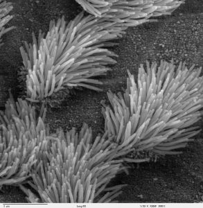
Cilia: Brush -like projections of the plasma membrane . Cilia for example can be found on human lung cells and they help to sweep foreign particles , inhaled dust, smoke and harmful microbes from entering the lungs. In fallopian tubes they move the ovum towards the uterus. Cilia generate water currents to carry food and oxygen past the gills of clams . They transport food through the digestive system of snails. Protozoans belonging to the phylum ciliophora are covered with cilia.
 “Cilia on Bronchiolar epithelium” by Charles Daghlian via Wikimedia Commons is in the Public Domain, CC0
Flagella : These are the whip-like extensions of the plasma membrane . They are found on gametes . They create the water currents necessary for respiration and circulation in sponges and coelenterates . Flagella are characteristic of the protozoan group Mastigophora

“Flagella” by Mike Jones, CC BY 3.0 , via Wikimedia Commons
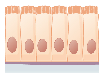
Microvilli: These are finger-like projections of the plasma membrane and are found in cells specialised in absorption . Such cells are found lining the small intestine, they help to absorbs nutrients from digested food.

“Microvilli” by OpenStax CNX is licensed under CC BY 4.0
Cytoplasm
The entire region of the cell between the plasma membrane and the nuclear envelope is filled filled with a jelly-like substance called the cytoplasm.
The cytoplasm provides the fluid medium necessary for biochemical reactions like Glycolysis, Protein synthesis .
The cytoplasm consists of 70to 80 percent water .
The semi-solid consistency of the cytosol is due to its protein content.
The other organic constituents of the cytoplasm includes Glucose and other simple sugars, polysaccharides, amino acids, nucleic acids, fatty acids, Glycerol and its derivatives.
Ions of sodium, potassium, calcium, and many other elements are also dissolved in the cytoplasm.
The Cytoskeleton
These are the network of protein fibers within the cytoplasm
They help to maintain the shape of the cell, hold certain organelles in specific positions, allows cytoplasm and vesicles to move within the cell, and enables unicellular organisms to move independently.
This network of protein fibers is collectively known as the cytoskeleton.
There are three types of fibers within the cytoskeleton: microfilaments, also known as actin filaments, intermediate filaments, and microtubules
Microfilaments : These are the thinnest of the cytoskeletal fibers . They help in moving cellular components, for example, during cell division. They also maintain the structure of microvilli. These components are also common in muscle cells and are responsible for muscle cell contraction.
Intermediate filaments : These are of intermediate diameter. They maintain the shape of the cell and anchor organelles .Example of intermediate filament is Keratin, the compound that strengthens hair and nails.
Microtubules : These are the thickest of the cytoskeletal fibers. These are hollow tubes . They can dissolve and reform quickly. They help in organelle movement. They pull chromosomes to their poles during cell division. They are also the structural components of flagella and cilia
In cilia and flagella these are are organized as a circle of nine double microtubules on the outside and two microtubules in the center .
Test your understanding
The plasma membrane, also called the cell membrane, is the membrane found in all cells that separates the interior of the cell from the outside environment. In bacterial and plant cells, a cell wall is attached to the plasma membrane on its outside surface
The centrosome is an organelle present in an animal cell that acts as the microtubule-organizing center of the cell. It also regulates the cell cycle
A lysosome is a membrane-bound cell organelle that contains digestive enzymes. Lysosomes are involved in various cell processes like break down excess or worn-out cell parts, destroying invading viruses and bacteria,programmed cell death, or apoptosis.
Brush-like projections of plasma membrane
Whip-like extensions of the plasma membrane
Finger-like projections of plasma membrane . They help in absorption
These are thin cytoskeletal fibres and they help in cellular components movements
These are thickest cytoskeletal fibres ,they help in organelle movement . The pull the chromosomes to their poles during cell division


